39 chlamydomonas diagram with labels
Modular, cascade-like transcriptional program of regeneration in ... 04.08.2022 · In Chlamydomonas, radial spoke protein synthesis reaches its maximum rate 30–60 min after the flagella have begun assembling onto pre-existing basal bodies (Remillard and Witman, 1982). This timing roughly matches the delay of 1 hr seen in our data between the peak expression of genes involved in ciliary assembly (cluster 2) and genes encoding radial spokes … Structure of Chlamydomonas (With Diagram) | Chlorophyta In this article we will discuss about the structure of chlamydomonas with the help of suitable diagrams. Chlamydomonas is unicellular, motile green algae. The thallus is represented by a single cell. It is about 20 p,-30|i in length and 20 µ in diameter. The shape of thallus can be oval, spherical, oblong, ellipsoidal or pyriform.
Microorganisms: Friend and Foe Class 8 Extra Questions ... Oct 11, 2019 · Pull out a gram or bean plant from the field. Observe its roots. You will find round struc¬tures called root nodules on the roots. Draw a diagram of the root and show the root nod¬ules. Answer: Question 2. Collect the labels from the bottles of jams and jellie on the labels. Answer: Do it yourself. Question 3. Visit a dcotor.

Chlamydomonas diagram with labels
Schematic representation of Chlamydomonas at different ... Download scientific diagram | Schematic representation of Chlamydomonas at different cell stages.-(A) Cell structure at biflagellate cell. Animal Cells: Labelled Diagram, Definitions, and Structure - Research Tweet Only present in lower plant forms (e.g. chlamydomonas) Present in all animal cells: Chloroplast: Plant cells have chloroplasts to synthesize their own food. Absent: Plasma Membrane: Cell wall and a cell membrane: Only cell membrane: Flagella: Present in some cells (e.g. sperm of bryophytes and pteridophytes, cycads and Ginkgo) The Chlamydomonas Flagellum as a Model for Human Ciliary Disease (The images and diagrams of Chlamydomonas central pairs are from Lechtreck and Witman, 2007; ... Although the antibody labeled internal membranes, the strongest labeling, and the only labeling on the surface of the cell, was in the cilium. These results demonstrated that polycystin-2 is displayed specifically on the primary cilium.
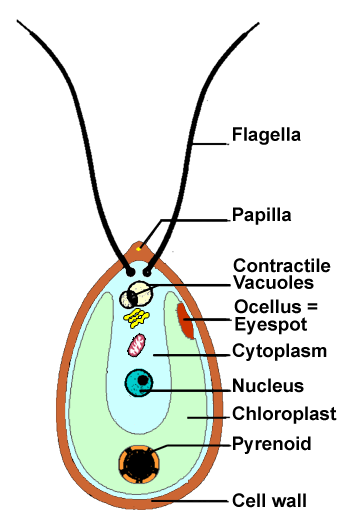
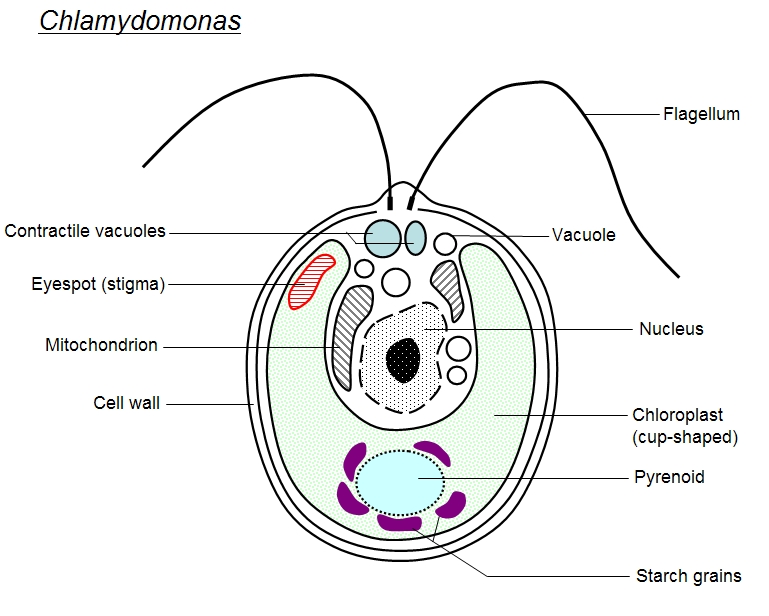
Chlamydomonas diagram with labels. Chlamydomonas - Meaning, Structure, Life Cycle, Function and FAQs - VEDANTU Every flagellum has two contractile vacuoles at the base. A small red eyespot can be found on the chloroplast's anterior side. Given below is the Chlamydomonas structure with labels. The Life Cycle of Chlamydomonas . Chlamydomonas Reproduction is both sexual as well as asexual reproduction. Asexual reproduction takes place by following methods: 1. Amoeba Diagram Illustrations, Royalty-Free Vector Graphics ... - iStock Browse 65 amoeba diagram stock illustrations and vector graphics available royalty-free, or start a new search to explore more great stock images and vector art. Newest results. Anatomy of an amoeba. Amoeba unicellular animal with pseudopods that lives in fresh or saltwater. Anatomy of an amoeba. Labeled Diagram of Spirogyra - QS Study Labeled Diagram of Spirogyra. Plant kingdom. Spirogyra is a sophisticated, filamentous green alga, found in freshwater represented by about 300 species. It is also identified as pond silk, as its fiber burnishes like silk due to the occurrence of mucilage. Describe the structure of chlamydomonas with neat labelled diagram ... answeredOct 30, 2020by Naaji(56.8kpoints) selectedOct 30, 2020by Jaini Best answer 1. Chlamydomonas is a simple, unicellular, motile fresh water algae. They are oval, spherical or pyriform in shape. 2. The cell is surrounded by a thin and firm cell wall made of cellulose. 3. The cytoplasm is seen in between the cell membrane and the chloroplast. 4.
Lifestyle | Daily Life | News | The Sydney Morning Herald The latest Lifestyle | Daily Life news, tips, opinion and advice from The Sydney Morning Herald covering life and relationships, beauty, fashion, health & wellbeing Characterization techniques for nanoparticles: comparison and ... The size distribution of their particles depended on the pH of the culture medium. The Ag NP toxicity on the green alga Chlamydomonas acidophila was pH-dependent as shown by the cytotoxicity mediated through the induction of oxidative stress. 227. Pavlopoulou et al. monitored the synthesis of Pt NPs using pH-responsive microgel particles. SAXS ... Brigitte Zimmer Well Labelled Diagram Of Chlamydomonas. By Admin August 21, 2022 Post a Comment. Shipping a package with ups is easy, as you can print labels for boxes, paste them and even schedul…. Read more. Use this labeled diagram of a chlamydomonas cell to - Course Hero Use this labeled diagram of a Chlamydomonas cell to address the following two questions. 32. Which of the following statements correctly identifies aspects related to photosynthesis and/or respiration? 1. Acetyl CoA is most often found in G. 2. NADPH accumulates in C. 3. ATP is found in F. 4.
Morphology of Chlamydomonas (With Diagram) | Algae - Biology Discussion In this article we will discuss about the external morphology of chlamydomonas. Also learn about its Neuromotor Apparatus, Electron Micrograph, Palmella-Stage with suitable diagram. 1. The organism is an unicellular alga (Fig. 11). 2. The thallus is spherical to oblong in shape but some species are pyriform or ovoid. ADVERTISEMENTS: 3. Biological drawings. Structure of Chlamydomonas. ... Dec 27, 2012 - Biological drawings of Protista, Structure of Chlamydomonas, Resources for Biology Education by D G Mackean. Learn CBSE - Microorganisms: Friend and Foe Class 8 Extra … 11.10.2019 · Pull out a gram or bean plant from the field. Observe its roots. You will find round struc¬tures called root nodules on the roots. Draw a diagram of the root and show the root nod¬ules. Answer: Question 2. Collect the labels from the bottles of jams and jellie on the labels. Answer: Do it yourself. Question 3. Visit a dcotor. Draw a neat labelled diagram. Chlamydomonas - Biology Draw a neat labelled diagram. Chlamydomonas . Maharashtra State Board HSC Science (General) 11th. Textbook Solutions 8018. Important Solutions 19. Question Bank Solutions 5546. Concept Notes & Videos 439. Syllabus. Advertisement Remove all ads. Draw a neat labelled diagram. ...
[Groenwieren: Green algae: Chlorophytae
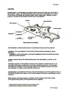
Biological drawings. Structure of Chlamydomonas. Learning Resources for ... Structure of Chlamydomonas: Next Drawing > Chlamydomonas is the name given to a genus of microscopic, unicellular green plants (algae) which live in fresh water. Typically their single-cell body is approximately spherical, about 0.02 mm across, with a cell wall surrounding the cytoplasm and a central nucleus.

Chlamydomonas Diagram With Labels
Structure and Diagram of Volvox and Their Functions - NotesHippo Volvox Structure: Diagram of Volvox with Label The cells of anterior end possess bigger eye spots than those of posterior end cells. The cells of posterior side become reproductive on maturity. Thus, spherical or round colony of Volvox shows clear polarity. Cell structure of volvox colony are Chlamydomonas type.

33 best Protists images on Pinterest | Ap biology, Biology lessons and Life science
Substancial | PDF | United Kingdom | Spain - Scribd substancial - Free ebook download as Text File (.txt), PDF File (.pdf) or read book online for free. contains some random words for machine learning natural language processing

Untitled | Pearltrees
Chlamydomonas reinhardtii growth is normal in TAP and 15 N-TAP but... Chlamydomonas reinhardtii growth is normal in TAP and 15 N-TAP but impaired in media containing D2O. Algal cell cultures were grown in each medium TAP, 15N-TAP and TAP containing each of the...
Chlamydomonas | ClipArt ETC
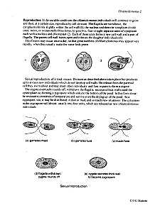
Life Cycle of Chlamydomonas (With Diagram) - Biology Discussion Each daughter cell develops cell wall, flagella and transforms into zoospore (Fig. 6). The zoospores are liberated from the parent cell or zoosporangium by gelatinization or rupture of the cell wall. The zoospores are identical to the parent cell in structure but smaller in size. The zoospores simply enlarge to become mature Chlamydomonas.
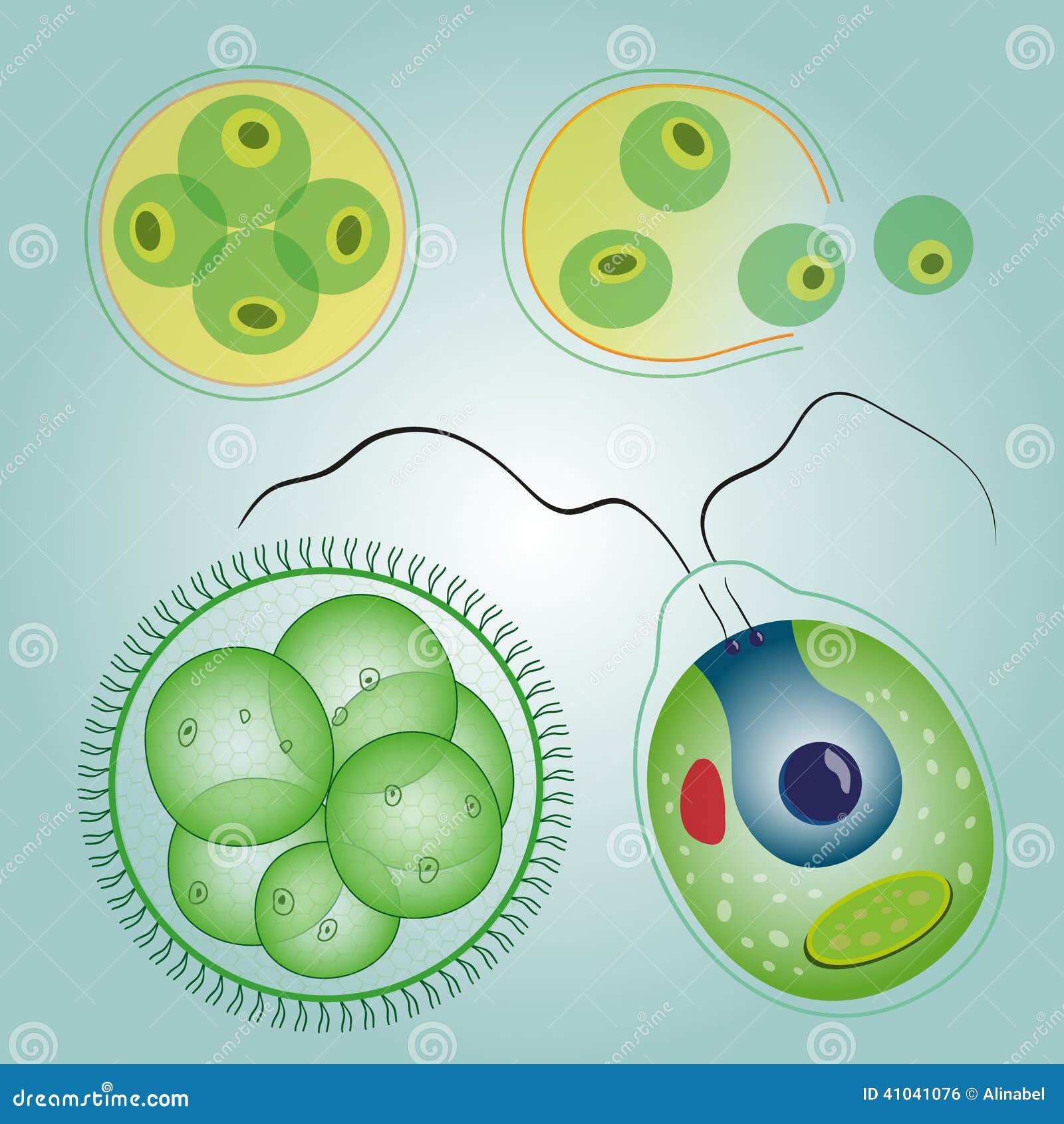
Algae Unicellular: Volvox, Chlorella And Chlamydomonas Stock Vector - Image: 41041076
MIT - Massachusetts Institute of Technology a aa aaa aaaa aaacn aaah aaai aaas aab aabb aac aacc aace aachen aacom aacs aacsb aad aadvantage aae aaf aafp aag aah aai aaj aal aalborg aalib aaliyah aall aalto aam ...
Algae
MIT - Massachusetts Institute of Technology a aa aaa aaaa aaacn aaah aaai aaas aab aabb aac aacc aace aachen aacom aacs aacsb aad aadvantage aae aaf aafp aag aah aai aaj aal aalborg aalib aaliyah aall aalto aam ...

Labelled Diagram Of Chlamydomonas - Top Label Maker
Draw a labelled diagram of Chlamydomonas. - Brainly.in 6 Oct 2019 — This is Expert Verified Answer · Chlamydomonas is a unicellular, motile freshwater species belonging to the genus of green algae. · They are oval, ...

207 best Algae images on Pinterest | Microbiology, Seaweed and Cell biology
Structure of Chlamydomonas (With Diagram) | Genetics - Biology Discussion In this article we will discuss about the structure of chlamydomonas (explained with labelled diagram). The unicellular green alga Chlamydomonas is haploid with a single nucleus, a chloroplast and several mitochondria (Fig. 9.3). It can reproduce asexually as well as sexually by fusion of gametes of opposite mating types (mt + and mt - ).
Structure Used By Amoeba For Movement
Amoeba Diagram Pictures, Images and Stock Photos Browse 116 amoeba diagram stock photos and images available, or start a new search to explore more stock photos and images. Anatomy of an amoeba. Amoeba unicellular animal with pseudopods that lives in fresh or saltwater. Anatomy of an amoeba. Vector illustration for medical, educational and science use. Amoeba labeled vector illustration.
Chlamydomonas Diagram With Labels
Higher Education Support | McGraw Hill Higher Education Learn more about McGraw-Hill products and services, get support, request permissions, and more.

Chlamydomonas Diagram With Labels
Chlamydomonas | Facts, Structure, Life Cycle, & Classification Chlamydomonas, genus of biflagellated single-celled green algae (family Chlamydomonadaceae) found in soil, ponds, and ditches. Chlamydomonas species can become so abundant as to colour fresh water green, and one species, C. nivalis, contains a red pigment known as hematochrome, which sometimes imparts a red colour to melting snow. The cells of most Chlamydomonas species are more or less oval ...

User:Nicole Bonan/Notebook/Biology 210 at AU - OpenWetWare
Substancial | PDF | United Kingdom | Spain - Scribd substancial - Free ebook download as Text File (.txt), PDF File (.pdf) or read book online for free. contains some random words for machine learning natural language processing

Chlamydomonas | ClipArt ETC
Chlamydomonas: Position, Occurrence and Structure (With Diagrams) Chlamydomonas is unicellular, motile green algae. The thallus is represented by a single cell. It is about 20 p,-30|i in length and 20 µ in diameter. The shape of thallus can be oval, spherical, oblong, ellipsoidal or pyriform. The pyriform or pear shaped thalli are common, they have narrow anterior end and a broad posterior end (Fig. 1).
Post a Comment for "39 chlamydomonas diagram with labels"